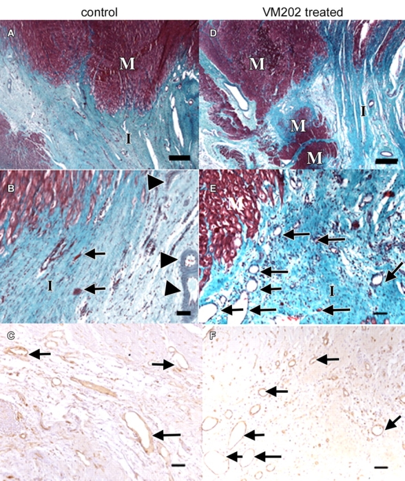
Figure 7:

Light microscopic photographs of transverse slices of control and VM202-treated hearts. A, At low magnification, there is a sharp boundary between the scar of the healed infarct (I) and viable myocytes (M) in control heart 7–8 weeks after coronary artery occlusion and reperfusion. B, At the edge of the same infarction at higher magnification, few sparse thin-walled vessels (arrows) and thick-walled vessels (arrowheads) are present, as well as degenerated myocytes. C, Blood vessels (arrows) in periinfarcted myocardium are localized with brown reaction product of lectin stain. D, In contrast, a treated heart at low magnification shows irregular healed infarct (I) borders with several peninsulas and/or islands of viable myocytes (M) within it. E, At high magnification, the edge of the scar contains numerous thin-walled blood vessels (arrows in E and F). Calibration bars in A and D = 150 μm. Calibration bars in B, C, E, and F = 45 μm. (A, B, D, and E, Masson trichrome stain; C and F, biotinylated isolectin B4 stain.)
