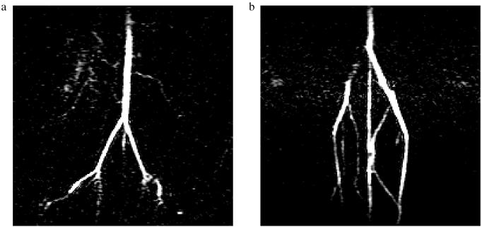
Figure 3.
In vivo non–slice-selective coronal images (0.31 × 0.31 mm resolution, 80 ms TR, 20° α) of rat vasculature acquired using 3He microbubbles suspended in Hexabrix. The imaging region of a is several centimeters cranial to the location of b. In a, the abdominal aorta, common iliac, and external iliac arteries are observable, and in b, the vena cava, common iliac, and caudal veins are visible.

