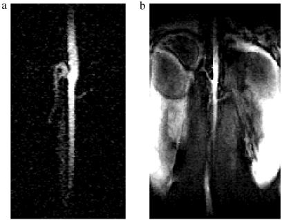
Figure 4.
(a) In vivo non–slice-selective coronal image of the renal arteries in a rat acquired using 3He microbubbles suspended in Hexabrix (0.31 × 0.31 mm resolution, 200 ms TR, 15° α). The following vessels are present: abdominal aorta, superior mesenteric artery, right and left renal arteries, and vena cava. (b) A 4.9-mm coronal section (seven slices) from a three-dimensional 1H image of the same location (0.31 × 0.31 mm in-plane resolution).

