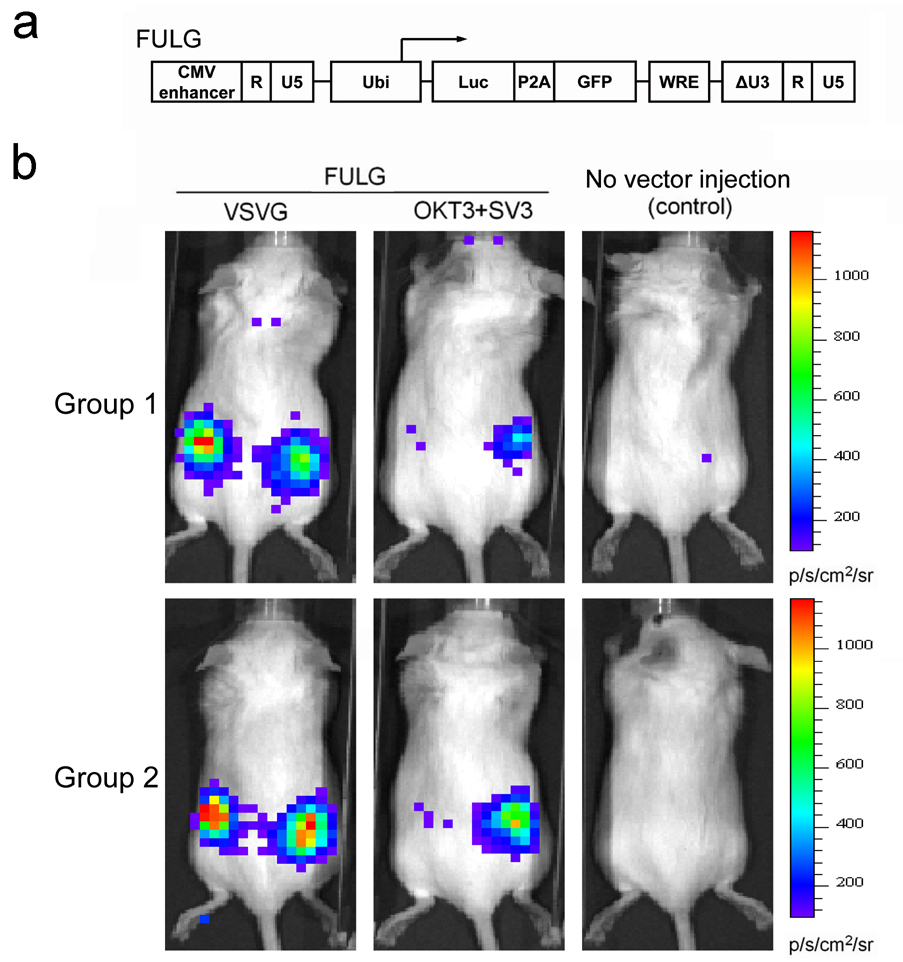
Fig. 8.
Targeting Jurkat.CD3 cells in vivo using engineered lentiviral vectors. (a) Schematic representation of a biocistronic lentiviral backbone plasmid capable of delivering two reporter genes. Luc: firefly luciferase gene; P2A: a self-cleavage peptide sequence derived from Porcine teschovirus; GFP: green fluorescence protein. (b) Targeted delivery of reporter genes in vivo in a xenografted mouse model. Jurkat.CD3 cells (5 × 10⁶) were injected subcutaneously on the right flank side of each mouse (RAG2−/−γc−/− female mice, 6–8 weeks). After 8 hours, the concentrated lentiviral vector (FULG/OKT3+SV3, 25 × 106 TU) was injected subcutaneously on both sides of each mouse. Six days later, firefly luciferase expression was imaged using a bioluminescence imaging system. FULG/VSVG was used as a non-targeting control. Two groups of experiments were conducted independently.

