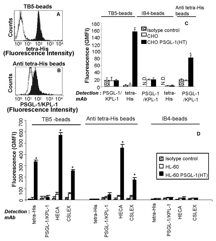
Figure 3.
Cytometry-bead experiments used to detect N-terminal PSGL-1(HT) peptide expressed in CHO and HL-60 cells. CHO/HL-60 PSGL-1 (HT) or wild-type CHO/HL-60 cells were treated with 50U/mL bovine thrombin in 200μl volume for 2h at 37°C. Supernatant containing cleaved peptide was collected and this was allowed to bind either TB5-beads that bear anti-PSGL-1 mAb (panels A) or anti tetra-His beads (panel B). PSGL-1 binding to these beads was detected using either anti-tetra-His mAb for panel A or anti-PSGL-1 mAb KPL-1 for B. Representative cytometry histogram are presented for CHO cells where black-filled peaks correspond to supernatant from cells expressing PSGL-1 (HT), grey-empty peaks correspond to wild type cells, and black-empty peaks represent isotype control sample. Panels C and D present a summary of the cytometry data for CHO and HL-60 cells respectively as mean + SEM (N=3). In both panels, immunoprecipitated PSGL-1 fragment is detected using both TB5- and anti tetra-His beads, but not isotype IB4-beads. Also, the binding sites for clones TB5 and KPL-1 overlap. Glycans on the PSGL-1(HT) peptide from HL-60 is detected using both anti-CLA mAb HECA-452 and anti-CD15s mAb CSLEX-1, on both TB5- and Anti tetra-His beads. N.D.: Not done since these are isotype controls themselves. *p<0.05 with respect to wild-type CHO/HL-60 cells.

