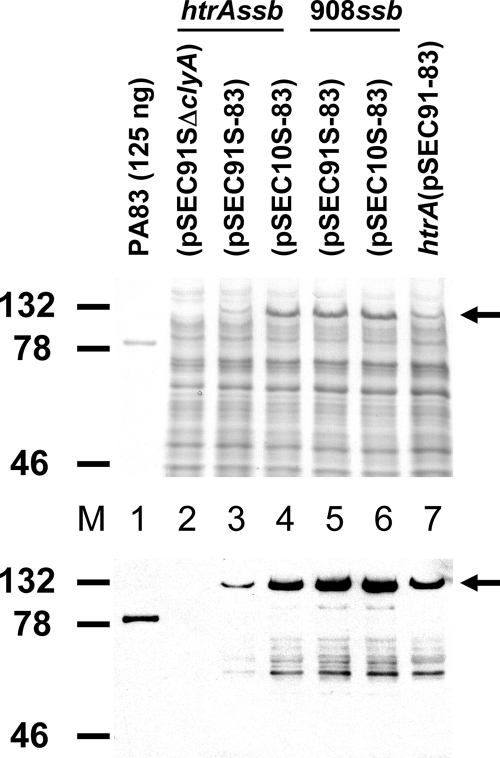
FIG. 4.
Coomassie brilliant blue-stained (top) and Western immunoblot analysis (bottom) of whole bacterial lysates from live vectors expressing ClyA-PA83 fusions from SSB-maintained plasmids carried by CVD 908-htrAssb (lanes 2 to 4) and CVD 908ssb (lanes 5 and 6). Lysates from strains carrying medium-copy-number derivatives of pSEC91S are in lanes 2, 3, and 5; lysates from strains carrying low-copy-number derivatives of pSEC10S are in lanes 4 and 6. A lysate from the conventional strain CVD 908-htrA(pSEC91-83) expressing ClyA-PA83 from a medium-copy-number kanamycin resistance plasmid is in lane 7, and 125 ng of purified PA83 is in lane 1. Total protein from approximately 106 CFU was resolved in each lane. Detection of ClyA-PA83 fusions was carried out using a polyclonal goat anti-PA83 primary antibody. The values at the left are molecular masses in kilodaltons. The arrows on the right indicate the position of the ClyA-PA83 fusion protein, with an expected molecular mass of 117 kDa.

