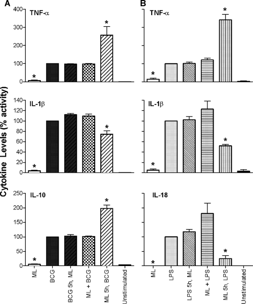
FIG. 5.
Cytokine response of monocytes costimulated with M. leprae, M. bovis BCG, and LPS. Monocytes from healthy human donors were stimulated with M. leprae (ML) or BCG alone or with ML and BCG simultaneously or were preexposed to ML or BCG for 5 h, followed by stimulation with the other organism (A); alternatively, monocytes were stimulated with ML or LPS alone or with ML and LPS simultaneously or were preexposed to ML or LPS for 5 h, followed by exposure to the other stimulant (B). Cell supernatants were collected at 48 h postinfection, and cytokines were analyzed by Luminex assay. Values are expressed as percent activity over BCG-stimulated (A) or LPS-stimulated (B) cells. Results are the means ± standard error of four experiments (representing four independent donors) performed in duplicate. A two-tailed, paired t test was used for statistical analysis (*, P < 0.05, in comparison to BCG- or LPS-stimulated cells).

