FIG. 4.
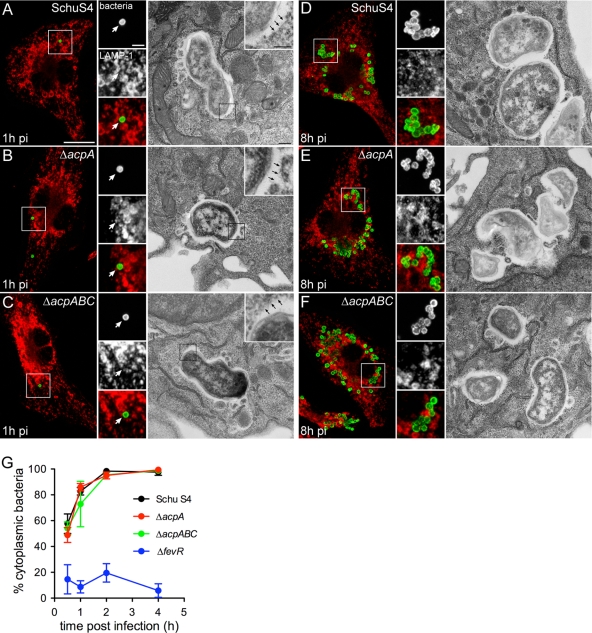
Intracellular trafficking of Schu S4 is not affected by deletion of the acp genes. muBMMs were infected with strain Schu S4, Schu S4 ΔacpA, or Schu S4 ΔacpABC and were processed for either immunofluorescence or transmission electron microscopy. (A to F) Representative confocal fluorescence or electron micrographs of muBMMs at 1 h (A to C) or 8 h (D to F) after infection with either Schu S4 (A and D), Schu S4 ΔacpA (B and E), or Schu S4 ΔacpABC (C and F). Bacteria appear green; LAMP-1 appears red. White arrows on the confocal micrograph insets indicate bacteria that are not surrounded by LAMP-1-positive membranes. Black arrows on the electron micrograph insets indicate a lack of phagosomal membranes. Bars, 10 and 2 μm for confocal microscopy panels and insets, respectively; 0.5 and 0.2 μm for electron microscopy panels and insets, respectively. (G) Phagosomal escape kinetics of strains Schu S4, Schu S4 ΔacpA, Schu S4 ΔacpABC, and Schu S4 ΔfevR in muBMMs. Macrophages were infected with individual strains, and were processed for a phagosomal integrity assay, as described in Materials and Methods. Values are means ± standard deviations for three independent experiments.

