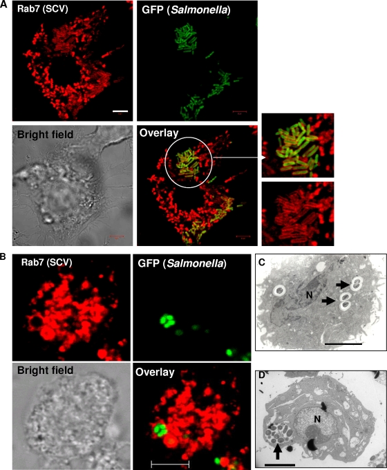
FIG. 1.
SCVs contain a single bacterium per vacuole. (A) Confocal laser scanning microscope images of RAW 264.7 cells infected with GFP-expressing Salmonella for 12 h and immunostained for Rab7 (red). The image shows many SCVs that have a single bacterium per vacuole. The white circle indicates the enlarged part. (B) Micrographs of an SCV containing multiple bacteria. RAW 264.7 cells were treated with 100 μM of sodium orthovanadate for 3 h, after which they were infected with GFP-expressing Salmonella for 12 h and immunostained for Rab7 (red), and the image was taken using a confocal laser scanning microscope. Sodium orthovanadate was maintained throughout the experiment. (C) Transmission electron microscope image of a RAW 264.7 cell infected with Salmonella for 12 h. The image shows SCVs that have a single bacterium per vacuole and also demonstrates the division of SCVs along with the bacteria (arrows). (D) Transmission electron microscope image of a RAW 264.7 cell treated with sodium orthovanadate (100 μM) and infected with Salmonella as described for panel B. The image shows an SCV that has multiple bacteria (arrow). N, nucleus. In all images, the scale bars correspond to 5 μm.

