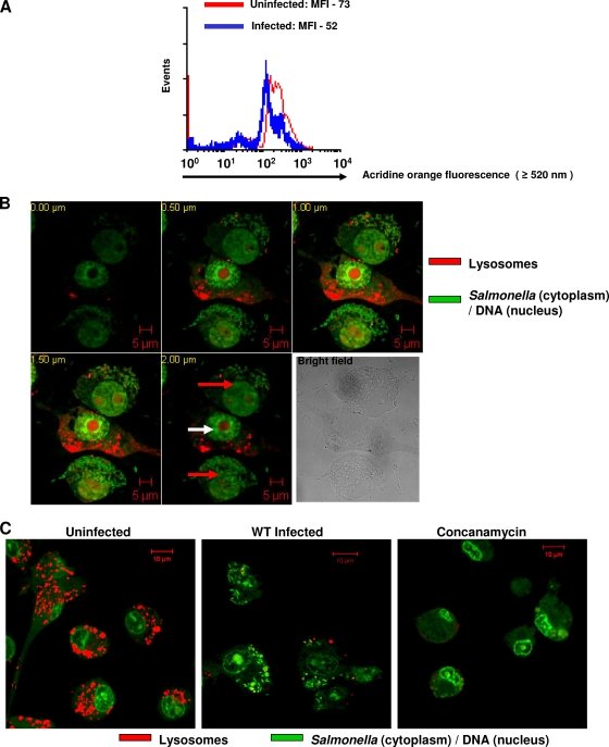
FIG. 6.
Salmonella infection reduces the volume of acidic compartments contributed by lysosomes in murine macrophages as inferred from acridine orange fluorescence. (A) Flow cytometric analysis of RAW 264.7 cells infected with Salmonella (MOI, 50 for 10 h) and stained with acridine orange. The difference in MOIs is statistically significant (P < 0.05; Student's t test). MFI, mean fluorescence intensity. (B) Confocal laser scanning microscope images of RAW 264.7 cells infected with GFP-expressing Salmonella for 10 h and stained with acridine orange. Five different optical sections (0.5-μm interval) of three RAW 264.7 cells are shown. The upper and lower cells are infected (red arrows), whereas the middle cell is uninfected (white arrow). (C) Confocal laser scanning microscope images of RAW 264.7 cells stained with acridine orange. Representative images of cells either uninfected or infected with GFP-expressing Salmonella or treated with 50 nM of concanamycin A are shown. Rod-shaped GFP-expressing Salmonella bacteria can be seen inside infected cells in both panels B and C. The results are representative of at least two independent experiments. Scale bars, 5 μm (B) and 10 μm (C).

