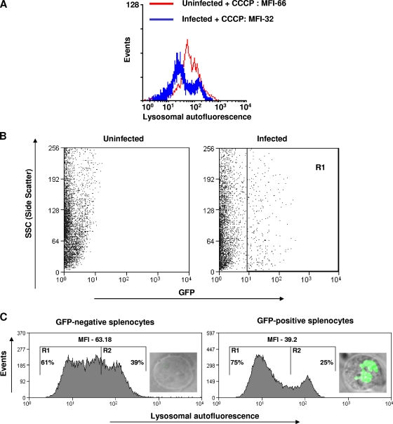
FIG. 7.
Salmonella reduces the lysosomal autofluorescence of host cells. (A) Flow cytometric analysis of lysosome-specific autofluorescence of RAW 264.7 cells infected with Salmonella (MOI, 10 for 10 h). Ten micromolar of carbonyl cyanide m-chlorophenylhydrazone was used to block the mitochondrial autofluorescence. The difference in MFI is statistically significant (P < 0.05; Student's t test). (B) Flow cytometry profile of splenocytes isolated from the spleens of either uninfected mice or mice infected intraperitoneally with GFP-expressing Salmonella (104 bacteria per mouse). GFP-positive cells (region R1) and GFP-negative cells were sorted using a fluorescence-assisted cell sorter. (C) Lysosome-specific autofluorescence of GFP-positive and GFP-negative splenocytes (20,000 each) isolated from a Salmonella-infected mouse. Ten micromoles of carbonyl cyanide m-chlorophenylhydrazone was used to block the mitochondrial autofluorescence. The difference in MFI is statistically significant (P < 0.05; Student's t test). The percentages of cells falling in R1 or R2 are shown. Representative images of GFP-positive (infected) and GFP-negative (uninfected) splenocytes are shown inside the respective graphs. The results are representative of two independent experiments.

