FIG. 1.
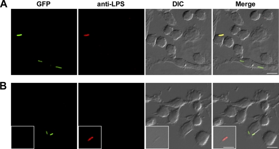
T6SS-1 is expressed following uptake of B. mallei by RAW 264.7 macrophages. Monolayers infected with B. mallei BM0739G (pBHR2-virAG) or BM0739G (pBHR1) were fixed at 3 h postinfection, immunostained under nonpermeabilizing conditions, and examined by DIC and fluorescence microscopy. GFP-expressing bacteria are shown in green while extracellular bacteria stained with the 3D11 MAb are shown in red. (A) Constitutive expression of GFP by B. mallei BM0739G (pBHR2-virAG). (B) Inducible expression of GFP by BM0739G (pBHR1) following uptake into RAW 264.7 cells; inset demonstrates inability of extracellular bacteria to express GFP. Micrographs are representative of at least three independent experiments. Scale bar, 10 μm.

