FIG. 7.
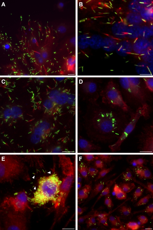
B. mallei tssE mutants demonstrate actin motility and intercellular spread defects in RAW 264.7 cells. Monolayers infected with B. mallei SR1A (pBHR1-TG) (A and B), BM0739G (pA0739G) (C) or BM0739G (pBHR1) (D, E, and F) were fixed at 24 h postinfection, stained, and examined by fluorescence microscopy. For panels A to E, monolayers were infected at an MOI of 20. For panel F, monolayers were infected at an MOI of 200. Bacteria expressing GFP are shown in green, host cell actin stained with Alexa Fluor 568-phalloidin is shown in red, and nuclei stained with DRAQ5 are shown in blue. White arrowheads in panel E indicate evidence of actin tail formation and potential intercellular spread. Micrographs are representative of at least three independent experiments. Scale bar, 10 μm.

