
FIG. 2.
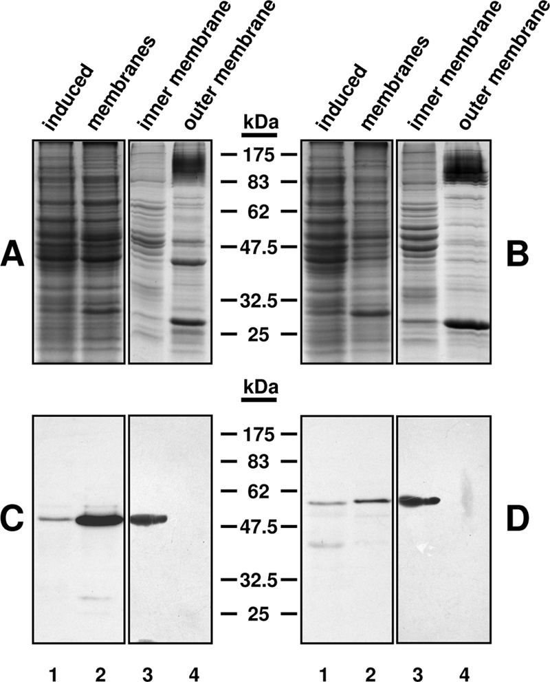
Subcellular localization of Lpt3 and Lpt6. Induced cell culture lysates (lanes 1) were clarified by centrifugation to remove inclusion bodies, followed by isolation of complete membrane fractions by ultracentrifugation (lanes 2), which were further separated into inner membrane (lanes 3) and outer membrane (lanes 4) fractions by sucrose gradient centrifugation. (A and B) Coomassie-stained 12.5% SDS-polyacrylamide gel of fractionation of lysates from induced cultures of the Lpt3-His6 expression strain CQWJR1 (A) and the Lpt6-His6 expression strain CQWJR2 (B). (C and D) Western immunoblot of the samples in panels A and B using monoclonal anti-polyhistidine peroxidase conjugate clone His-1 (Sigma Aldrich). For inner and outer membrane fractions, 10 μg total protein was loaded per lane. Note that both Lpt3-His6 and Lpt6-His6 localize to the inner membrane fraction.