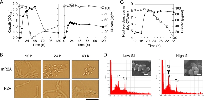
FIG. 2.
Silicate uptake during growth of B. cereus YH64. (A) Growth of YH64 (closed circles) on mR2A (left) and R2A (right) media containing 100 μg/ml silicate and silicate concentrations in the medium (open squares) were measured at different time points. (B) Phase-contrast microscopic images of YH64 that grew on mR2A and R2A media. Scale bar, 10 μm. (C) Silicate concentrations (open squares) and the numbers of heat-resistant spores (closed triangles) of YH64 culture containing 100 μg/ml silicate. The cell suspension was heated at 65°C for 30 min, and the number of heat-resistant spores that formed colonies on an R2A agar plate was determined. (D) EDX spectrum of YH64 spores. The EDX signal of silicon is indicated by an arrowhead. Insets are the SEM images of the spores. Scale bars, 1 μm.

