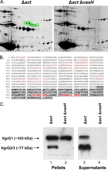
FIG. 1.
Identification of proteins secreted via T6SS in the supernatant of A. hydrophila SSU. (A) Comparison of 2-D gels containing proteins from the supernatants of the A. hydrophila SSU Δact (left) and Δact ΔvasH (right) mutant strains. Highlighted spots (in green) represent a cluster of proteins secreted via the T6SS which were identified by mass spectrometric analysis. (B) Alignment of VgrG1 from A. hydrophila ATCC 7966 (gi|117619461) with peptides identified via mass spectrometry (red) in one of the secreted proteins (VgrG1) from A. hydrophila SSU. The bold, underlined sequence represents the VIP-2 domain. (C) Western blot analysis of pellet and supernatant fractions of A. hydrophila SSU Δact (lanes 1 and 3) and Δact ΔvasH (lanes 2 and 4) mutant strains using antibodies specific to the VIP-2 domain of VgrG1 in combination with specific antibodies against VgrG2.

