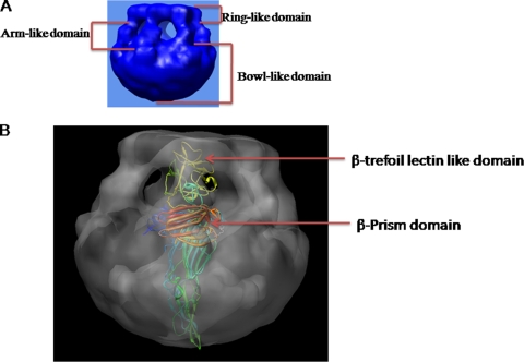
FIG. 9.
Model of pore-forming mechanism of 65-kDa hemolysin oligomer. (A) There are three main parts of the 3D structure of the 65-kDa HlyA oligomer. The top portion is the ring-like domain, the middle portion is the arm-like domain, and the bottom portion is the bowl-like domain. (B) X-ray crystallography structure fitted into the electron density map of the HlyA oligomer (side view). The β-trefoil lectin-like domain and the β-prism lectin are marked by arrows. The β-prism lectin domain is located in the junction of the arm-like and bowl-like domains in the 3D structure of the HlyA oligomer. The β-trefoil lectin-like domain is situated at the ring-like domain in the 3D structure of the HlyA oligomer.

