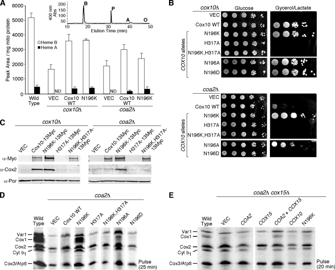
FIG. 4.
Catalytically active Cox10 and heme a generation are required for suppressor activity by N196K Cox10. (A) Heme was extracted from mitochondria (1.5 to 2 mg protein) isolated from WT cells, cox10Δ cells expressing YCp WT Cox10 or N196K Cox10, and coa2Δ cells expressing YCp WT Cox10 or Cox10-N196K and separated by reverse-phase high-performance liquid chromatography. The inset shows a representative chromatogram from WT mitochondria. The peaks corresponding to heme b (B), heme a (A), and protoporphyrin (P) and the expected elution time of heme o (O) are indicated. The white bars indicate the area under the heme b peak normalized to mg of mitochondrial protein, and the black bars represent the area under the heme a peak also normalized to mg of mitochondrial protein. The data represents the averages of three independent repeats, and the error bars represent the standard errors of the means. ND, not detectable. (B) cox10Δ and coa2Δ cells expressing WT Cox10, N196K Cox10, H317A Cox10, N196K-H317A Cox10, N196A Cox10, and N196D Cox10 were grown in SC-2% raffinose-0.2% glucose selective medium, serially diluted, and drop tested on SC-2% glucose or yeast extract-peptone-2% glycerol-2% lactate. The plates were incubated at 30°C for 2 days (glucose) or 4 days (glycerol/lactate). (C) Purified mitochondria (30 μg protein) from cox10Δ and coa2Δ cells expressing WT Cox10-13Myc, N196K Cox10-13Myc, H317A Cox10-13Myc, and N196K-H317A Cox10-13Myc were separated by SDS-PAGE and analyzed by immunoblotting using anti-Myc, anti-Cox2, and anti-Por1 (loading control) antibodies. (D) In vivo mitochondrial translation. W303 coa2Δ cells expressing WT Cox10, N196K Cox10, H317A Cox10, N196K-H317A Cox10, N196A Cox10, and N196D Cox10 were pulsed with [35S]methionine as for Fig. 1B. (E) Analysis of mitochondrial translation products in W303 coa2Δ cox15Δ cells expressing COA2, COX15, WT COX10, and COX10 (N196K). The cells were pulsed with [35S]methionine for 20 min at 30°C and analyzed as for Fig. 1B.

