Figure 1.
Identification of a Novel Mutant of the Plant Endomembranes.
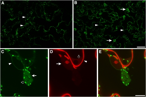
(A) and (B) Confocal microscopy images of live cotyledon epidermal cells of Arabidopsis of the control (nonmutagenized ST-GFP; [A]) and of the G92 mutant (B). Arrowheads show the presence of fluorescent spots that correspond to Golgi stacks. Note the presence of additional globular structures in the G92 sample (arrows in [B]) compared with the control (A). Bar = 50 μm.
(C) to (E) High-magnification confocal live-cell images of epidermal cells of the G92 (C) mutant treated with propidium iodide (D), which labels the cell wall ([D], arrowhead) and nucleic acids in the nucleoplasm ([D], arrow) and cytoplasm ([D], open arrow). The cell was imaged with confocal settings for GFP and propidium iodide (see Methods). Note the appearance of the fenestrated G92 globular structures ([C], arrow) that are close to the nucleus and that contain diffused and punctate ST-GFP fluorescence. In (C), an arrowhead and an open arrow point to the nuclear envelope that is also visible in the control (see Supplemental Figure 1 online) and to Golgi stacks, respectively. (E) shows merged images of (C) and (D). Bar = 10 μm.

