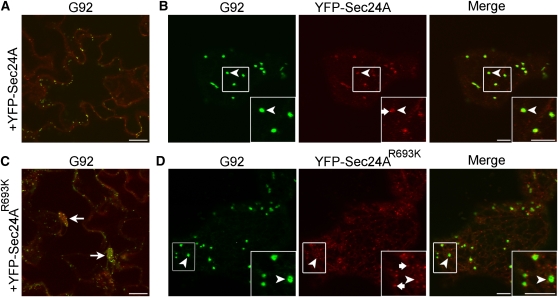
Figure 10.
Localization of YFP-Sec24A Is Affected by the R693K Mutation.
Confocal images of epidermal cells of cotyledons of T1 G92 plants expressing either 35S:YFP-Sec24 ([A] and [B]) or 35S:YFP-Sec24AR693K ([C] and [D]).
(A) The YFP-Sec24A construct complements the Sec24AR693K phenotype.
(B) At a higher magnification, YFP-Sec24 fluorescence is clearly visible at ERESs (main panels and insets, arrowheads) that are associated with the Golgi (main panels and insets, arrowheads). YFP-Sec24 fluorescence is also visible in the cytosol and at some small punctae (inset, arrow), consistent with previous reports (Stefano et al., 2006; Hanton et al., 2007, 2009).
(C) In contrast with YFP-Sec24A, YFP-Sec24AR693K did not complement the G92 mutant phenotype.
(D) Rather than being at Golgi-associated ERESs (main panels and insets, arrowheads), the fluorescence of YFP-Sec24AR693K appears to be distributed mostly at numerous small disperse punctae (inset, arrows). Insets show magnified sections of boxed regions in main images (2×). Bars in panels and insets = 5 μm.

