Figure 2.
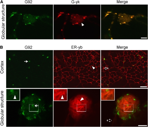
In Addition to Containing the trans-Golgi Marker ST-GFP, the G92 Structures Contain cis- and medial-Golgi Markers and Convoluted ER Tubules.
(A) Confocal live-cell images of a cotyledon epidermal cell of a G92 homozygous F2 plant expressing the cis/medial-Golgi marker G-yk. Note that the G-yk fluorescence in the globular structure (arrowhead) overlaps with that of ST-GFP. Bar = 5 μm.
(B) Cortex panels: Confocal images of the cortex and of a globular structure of a G92 cotyledon cell from a T1 seedling transformed with the ER marker ER-yb. Note the presence of a reticulated ER network of tubules with organization similar to that of a control cell (nonmutagenized ST-GFPx35S:ER-yk; see Supplemental Figure 4 online). Golgi stacks labeled with ST-GFP (G92 panel, arrow) lie on the ER tubules (arrowheads). The open arrow in the merge image shows one of the infrequent punctae that were labeled by ER-yb but not by ST-GFP in the G92 cotyledons. Globular structure panels: The G92 globular structure contains convoluted ER tubules (ER-yb panel, arrowhead) and Golgi (G92 panel, arrow; see also insets and Supplemental Figure 2 online). The ST-GFP fluorescence of the G92 mutant (G92 panel) overlaps with that of the ER marker in the globular structures (arrowheads). Insets show magnified sections of main panels (boxed area × 2). ER tubules extending to the G92 structure and labeled by ER-yb are indicated by an open arrow in (B) in the merged image. Bars in main images = 5 μm.

