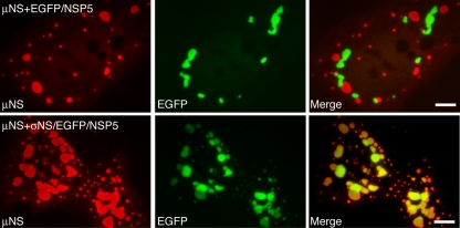
FIG. 1.
MRV μNS and EGFP-fused rotavirus NSP5 form nonoverlapping structures that can be made to colocalize through a known protein-protein association. CV-1 cells cotransfected with a plasmid expressing μNS and either EGFP/NSP5 (top) or σNS/EGFP/NSP5 (bottom) were fixed at 18 h p.t. FLS were visualized by staining with μNS-specific polyclonal antibodies followed by Alexa 594-conjugated goat anti-rabbit IgG (left). VLS were visualized by the inherent fluorescence of EGFP (middle). Merged images are also shown (right). Bar, 10 μm.

