FIG. 6.
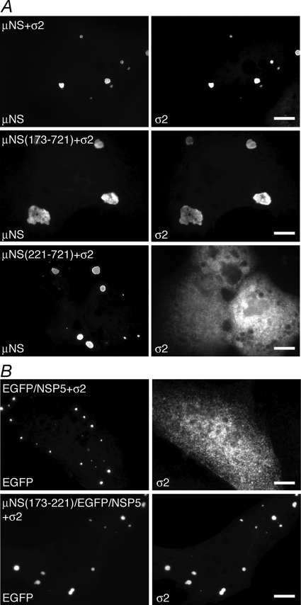
μNS aa 173 to 220 are necessary and aa 173 to 221 are sufficient for associations with the core surface protein σ2. For each experiment, cells were processed for fluorescence microscopy at 18 h p.t. (A) CV-1 cells were cotransfected with plasmids expressing σ2 and either μNS (top), μNS(173-721) (middle), or μNS(221-721) (bottom). After fixation, cells were stained with rabbit polyclonal antibodies against MRV cores followed by Texas Red-conjugated μNS-specific rabbit IgG to visualize μNS (left) and Alexa 488-conjugated goat anti-rabbit IgG to visualize σ2 (right). (B) CV-1 cells were cotransfected with plasmids expressing σ2 and either EGFP/NSP5 (top) or μNS(173-221)/EGFP/NSP5 (bottom). After fixation, cells were stained with rabbit polyclonal antibodies against MRV cores followed by Alexa 594-conjugated goat anti-rabbit IgG to visualize σ2 (right). The inherent fluorescence of EGFP was used to visualize each of the fusion proteins (left). Bar, 10 μm.

