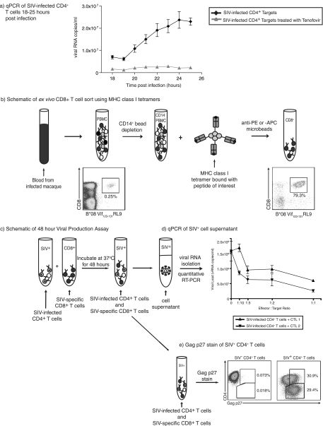
FIG. 1.
Viral life cycle timing and viral production assay schematic. (a) The length of the SIV life cycle was measured by analyzing viral RNA copies per milliliter of cell supernatant each hour between 18 and 25 h postinfection. Each assay for each time point was performed in triplicate. CD4+ T-cell targets were used from at least four animals and repeated in three different experiments. Cells pretreated with tenofovir for 2 h prior to infection and throughout the assay served as controls. (b) Effector cells were generated using MHC class I tetramers specific for the epitope of interest. Cells were incubated with either anti-PE or anti-APC Miltenyi microbeads depending on the tetramer fluorochrome used and passed through a Miltenyi magnetic column. (c) Activated, SIV-infected CD4+ T cells were incubated with ex vivo-sorted, unstimulated, SIV-specific CD8+ T cells for 48 h. (d and e) Supernatant was then removed for analysis of viral RNA content by quantitative RT-PCR (d), and the remaining cells were stained for CD4, CD8, and Gag p27 (e). qPCR, quantitative PCR.

