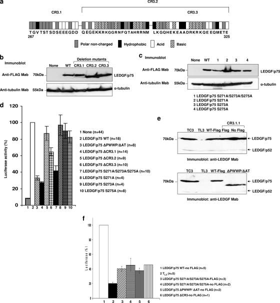
FIG. 3.
HIV-1 cofactor activity of different LEDGF/p75 CR3 mutants. (a) Analysis of charged residues in CR3 used to establish the boundaries of CR3.1, CR3.2, and CR3.3. (b and c) Expression of LEDGF/p75 mutants in TL3 cells evaluated by immunoblotting with an anti-FLAG MAb. Alpha-tubulin detection was used as a loading control. (d) Single-round infection of TL3 cells expressing LEDGF/p75 mutants. Cells evaluated in b and c were challenged with HIV-1 luciferase reporter viruses, and luciferase activity was analyzed 5 days later. Infectivity and error bars were calculated as described in the legend of Fig. 1d. (e) Immunoblotting detection of the reexpressed LEDGF/p75 WT and mutants in TL3 cells using an anti-LEDGF MAb. Detection of endogenous LEDGF/p52 was used as a loading control. (f) Single-round infection of TL3 cells expressing the nontagged LEDGF/p75 WT or mutants. Cells immunoblotted in e were challenged with HIVluc, and luciferase activity was analyzed 5 days later. Infectivity and error bars were calculated as described in the legend of Fig. 1d.

