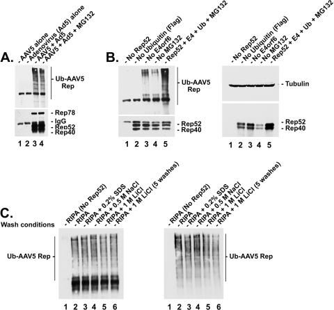
FIG. 1.
AAV5 Rep proteins are polyubiquitinated during coinfection with Ad. (A) Postimmunoprecipitation (IP) Western blot analysis of AAV5 Rep protein ubiquitination during AAV5-Ad coinfection. Western blots were performed as previously described (16). Briefly, cells were infected with AAV5 (MOI, 10) and/or Ad5 (MOI, 5) for 42 h. Where applicable, cells were treated after 36 h with either MG132 or equivalent amounts of dimethyl sulfoxide (DMSO) as a vehicle control. Cells were lysed with RIPA buffer containing 1% SDS and boiled to disassociate any interactions between Rep protein and other potentially ubiquitinated (Ub) cellular proteins. Lysates were diluted in RIPA buffer to a final SDS concentration of 0.2% and precleared once with protein G beads prior to immunoprecipitation. AAV5 Rep proteins were immunoprecipitated from 293-infected lysates with anti-Rep antibody and subjected to Western analysis using the anti-Rep (bottom) or anti-ubiquitin (top) antibodies. 293 cells were infected with AAV5 alone (lane 1), Ad5 alone (lane 2), or both AAV and Ad (lanes 3 and 4) in the absence (lane 3) or presence (lane 4) of the proteasome inhibitor MG132 (10 μM). The presence of ubiquitinated Rep proteins is indicated by the appearance of high-molecular-weight isoforms of the protein, observed as a laddering or smearing effect. (B) Western blot analysis of AAV5 small Rep protein ubiquitination in 293 cells in the absence or presence of adenovirus E4orf6 during transient transfection. Lysates were isolated 36 to 42 h posttransfection with vectors expressing AAV5 small Rep proteins plus a construct expressing E4orf6 and another expressing a Flag-tagged ubiquitin in the absence or presence of MG132 (added 6 h prior to cell harvest). Cells were transfected with each of the constructs except where noted: no Rep (lane 1), no Flag-ubiquitin (lane 2), no E4orf6 (lane 3), or no MG132 (lane 4). (Left panel) Post-IP Western blot following immunoprecipitation with an anti-Rep antibody, with an immunoblot using an anti-Rep (bottom) or anti-Flag (top) antibody. (Right panel) Pre-IP Western blot of the same samples as in the left panel, with anti-tubulin (top) or anti-Rep (bottom) antibodies. (C) Post-IP Western blot analysis of AAV5 small Rep protein ubiquitination in 293 cells in the presence of E4orf6 and MG132 during transient transfection following various stringent washing conditions. Cells were transfected with Flag-Ub and E4orf6 and treated with MG132 in the absence (lane 1) or presence (lanes 2 to 6) of AAV5 Rep52/40. Following immunoprecipitation, Rep/antibody-conjugated beads were washed twice with either RIPA buffer alone (lanes 1 and 2), RIPA buffer plus 0.2% SDS (lane 3), RIPA buffer plus 0.5 M NaCl (lane 4), or RIPA buffer plus 1 M LiCl (lane 5) or washed five times with RIPA buffer plus 1 M LiCl (lane 6). Samples were either blotted with anti-Rep antibody and exposed with high-sensitivity Femto reagent (left) or blotted with anti-ubiquitin antibody and exposed with Pico reagent (right). Antibody dilutions were as follows: anti-Flag (Sigma-Aldrich; #F1804), 1:5,000; anti-Rep (ARP; #03-61073), 1:200; anti-ubiquitin (Santa Cruz Biotechnology, Inc.; #sc-8017), 1:500; and goat anti-mouse horseradish peroxidase (HRP) conjugate (Sigma-Aldrich; #A5278) secondary antibody, 1:5,000.

