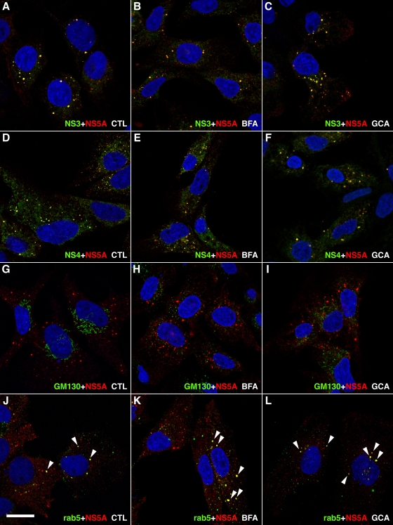
FIG. 6.
Immunofluorescence analysis of GBF1 inhibition in subgenomic replicon-harboring cells. Huh-7 cells harboring a subgenomic replicon were treated for 8 h in the presence of ethanol 0.2% (A, D, G, and J), 1 μg/ml BFA (B, E, H, and K), or 10 μM golgicide A (C, F, I, and L) and processed for double-label immunofluorescence using anti-NS5A antibody together with antibodies to NS3 (A to C), to NS4A-B (D to F), to cis-Golgi marker GM130 (G to I), or to Rab5 (J to L). Representative confocal images are shown, with NS5A in red and other markers in green. Colocalization of both markers in dot-like structures appears in yellow and is marked by white arrowheads in panels J to L. Bar, 20 μm.

