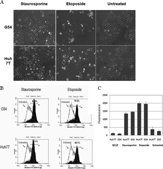
FIG. 2.
HCV replication does not suppress CDCA. CDCA was induced in both HCV-expressing G54 cells and the parental cells by treatment with the CDCA inducer staurosporine or etoposide. (A). Apoptosis, represented by cell morphology changes, was examined with a phase-contrast microscope. (B) Apoptotic cells were quantified by flow cytometry after being stained with annexin V-FITC. Shifting of the peak toward increased fluorescence (toward the right) represents apoptotic cells. The percentage of apoptotic cells is indicated. (C) The activity of caspase 3, a marker to determine CDCA activation, was assessed using a caspase 3 detection kit. CDCA was induced in G54 cells and parental Huh7T cells by treatment with staurosporine or etoposide. Caspase 3 activity was then detected with a fluorescence reader.

