FIG. 5.
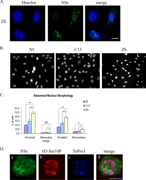
Infection with pathogenic RVFV strain ZH548 induces abnormal nuclear morphologies in kidney sheep cells. (A) Nonconfocal conventional fluorescence microscopy of kidney sheep cells of fetal origin infected by the RVFV ZH548 strain displaying total DNA distribution (Hoechst), NSs detected with anti-NSs antibody (green), and the corresponding merge image. (B) Nonconfocal conventional fluorescence microscopy of kidney sheep cells that were either not infected (NI) or infected by the RVFV ZH548 strain (ZH) or the nonpathogenic clone 13 (C13) strain displaying DNA stained with Hoechst. (C) The number of nuclei displaying abnormalities was quantified in uninfected (NI) and clone 13 (C13)- and ZH548 (ZH)-infected cells from two different experiments, with n > 800 at each condition. The incidence of abnormal nuclei in ZH-infected was compared to uninfected or C13-infected cells by using a chi-square test. *, P < 0.05; **, P < 0.01; ***, P < 0.001; NS, not significant. (D) Single confocal sections taken through the z axis of uninfected or ZH-infected mitotic kidney sheep cells displaying immunostaining with anti-NSs in green (a), immunostaining with anti-H3-Ser10P in red (b), DNA counterstained with ToPro3 in blue (c), and the corresponding merge images (d). Bars, 10 μm.

