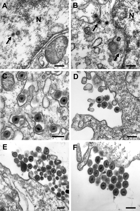
FIG. 6.
Transmission electron microscopy of cells infected with BHV1-ΔUL47, WT BHV-1, or BHV1-UL47R. MDBK cells were infected for 14 h with 5 PFU/cell of BHV1-ΔUL47 (A to D), WT BHV-1 (E), or BHV1-UL47R (F). (A and B) Nucleocapsids in the nucleus (N) and the cytoplasm (arrows), respectively, of the BHV1-ΔUL47-infected cells as evidence of unimpaired nuclear egress. (C) Apparently normal secondary envelopment of BHV1-ΔUL47 virions in the cytoplasm. (D to F) Very few extracellular viral particles were detected around the UL47-null virus-infected cells compared to the WT (E) or revertant (F) virus-infected cells, which were surrounded by numerous groups of extracellular virions. Bars, 200 nm.

