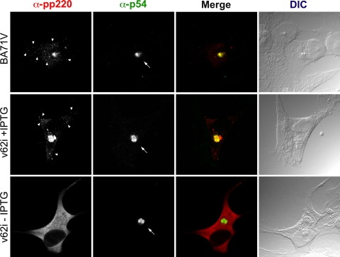
FIG. 5.
Immunofluorescence microscopy analysis of the subcellular localization of polyprotein pp220 in v62i-infected cells. Vero cells were fixed at 12 hpi with the parental BA71V or v62i in the presence or absence of 0.5 mM IPTG. Samples were incubated with the monoclonal antibody 18HH against the polyprotein pp220/p150 or with an antibody against the inner envelope protein p54, which was used as a marker for the viral factory. Antibodies were detected by using secondary antibodies coupled to Alexa 555 and Alexa 488, respectively. Differential interference contrast (DIC) microscopy of the samples is shown at right. Arrows and arrowheads indicate the positions of the viral factories and viral particles, respectively.

