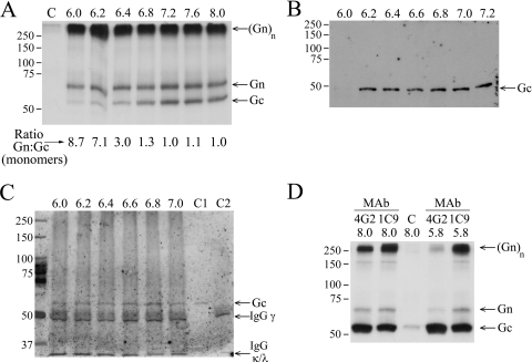
FIG. 1.
Effect of low-pH treatment on PUUV Gn-Gc complex in immunoprecipitation of virion lysates. (A) Gn-specific immunoprecipitation with MAb 5A2. Autoradiography of MAb 5A2-Sepharose-bound, [35S]cysteine-labeled Gn and Gc after washing at indicated pH values. The control lane C represents background binding to protein G Sepharose at pH 8.0 without MAb. SDS-PAGE was run under reducing conditions with 2-mercaptoethanol as the reducing agent. The band labeled (Gn)n may also contain Gc. (B) Loss of PUUV Gc recognition by Gc-specific MAb 4G2. Gc in nonlabeled PUUV lysate was bound to MAb 4G2-Sepharose in buffers at the indicated pH. Proteins were separated by reducing SDS-PAGE and immunoblotted with anti-Gc serum. (C) MAb 4G2 binding to Gc prevents dissociation at low pH. Nonlabeled PUUV lysate was bound to MAb 4G2-Sepharose at pH 8.0 and washed in buffers at the indicated pH. The material remaining bound was separated by reducing SDS-PAGE and immunoblotted with anti-Gc serum. C1 and C2 represent controls; C1 is a virus preparation used in immunoprecipitation, and C2 is MAb 4G2-Sepharose without addition of virus. IgG(γ) (∼50 kDa) and IgG(κ/λ) (25 kDa; migrating at front) chains are indicated. (D) The low-pH-induced dissociation of Gn-Gc complex is reversible. Lysates of metabolically ([35S]cysteine) labeled PUUV treated at pH 5.8 or kept at pH 8.0, as indicated, were immunoprecipitated with Gc-specific MAbs 4G2 and 1C9 at pH 8.0. Autoradiography of SDS-PAGE-separated proteins and the control (lane C) was performed as described for panel A. The protein mobility estimation in panels A and D was done using Precision Plus protein standards (Bio-Rad). The band labeled (Gn)n may also contain Gc.

