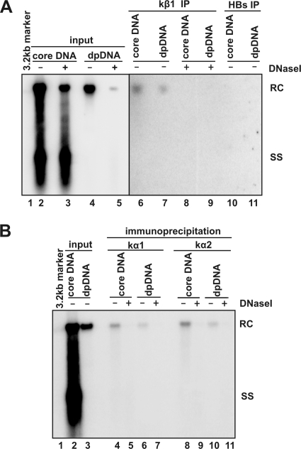
FIG. 6.
Cytoplasmic DP rcDNA were associated with karyopherins. One milliliter of cytoplasmic lysate prepared from 5 × 10⁵ HepDES19 cells that were cultured in the absence of tetracycline for 12 days was mixed with 35 μl of protein A/G plus beads (Santa Cruz) preabsorbed with antibodies against karyopherin-β1 (kβ1) (Abcam) or HBsAg (Dako) (panel A) and against karyopherin-α1 (kα1; clone 114-E12; Zymed) or karyopherin-α2 (kα2; sc-55537; Santa Cruz) (panel B). The mixtures were incubated at 4°C overnight. Beads were washed four times with TNE buffer, and core DNA and DP DNA were extracted from the beads with or without prior DNase I digestion as indicated. Viral DNA were analyzed by Southern blot hybridization. Lane 1 was loaded with 50 pg of 3.2-kb HBV DNA. In panel A, lanes 2 and 3 were loaded with 1/20 of core DNA from the cytoplasmic fraction of one 60-mm dish, and lanes 4 and 5 were loaded with half of DP DNA from the cytoplasmic fraction of one 60-mm dish. In panel B, lane 2 was loaded with 1/30 of core DNA from the cytoplasmic fraction of one 60-mm dish, and lane 3 was loaded with 1/10 of DP DNA from the cytoplasmic fraction of one 60-mm dish. Half the volume of immunoprecipitated core DNA and DP DNA recovered from one 60-mm dish was loaded onto the gel (panel A, lanes 6 to 11; panel B, lanes 4 to 11) The positions of rcDNA (RC) and single-stranded DNA (SS) are indicated.

