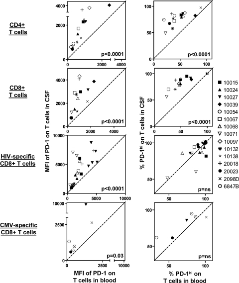
FIG. 2.
Increased PD-1 expression on T cells in CSF compared to those in blood from HIV-positive patients. (Left) Relationships between MFIs of PD-1 in blood and in CSF. (Right) Relationships between frequencies of PD-1hi T cells in blood and in CSF. Data from 15 HIV-positive patients are shown. On plots for CD4+ and CD8+ T cells, each data point represents an individual participant. On plots for HIV-specific and CMV-specific CD8+ T cells, each data point represents an individual tetramer response for each participant studied. The diagonal dashed line represents the line of unity. P values were derived from a two-tailed Wilcoxon signed-rank t test. Individual subject identifiers are listed to the right.

