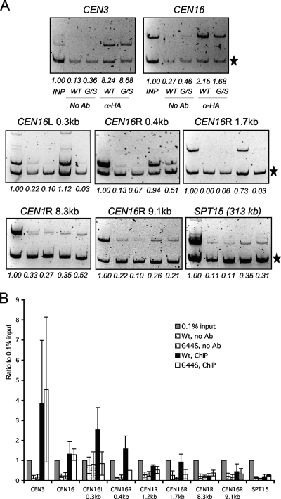
FIG. 6.
The G44S mutation selectively downregulates the pericentric recruitment of Sgo1p in vivo. (A) ChIP analysis of HA-tagged Sgo1p. Samples in all of the panels are arranged in the same order. The common internal control (marked by the star on the right) for multiplex PCRs is from within the ORF of the DED1 gene 386.2 kb to the right of CEN15. Targets of the PCR fragments and their distance to the cognate centromeres are listed at the top of each gel image. R, right; L, left. SPT15 is 313.3 kb to the right of CEN5. (B) Quantification of ChIP results. Ethidium bromide-stained DNA gel images were quantified by NIH Image. The intensity of each CEN or pericentric fragment was compared to that of the DED1 internal control (star). The ratio was then normalized to 0.1% input (INP) DNA (set at 1.0). Error bars represent standard deviations from at least three independent cell cultures for ChIP. Ab, antibody.

