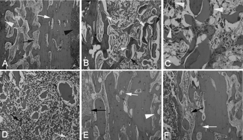
FIG. 6.
Representative photomicrographs of longitudinal sections of tibiae (hematoxylin and eosin stained). (A) Typical signs of osteomyelitis prior to surgery included destruction of bone (white arrow), reactive hyperostosis (white arrowhead), fibrosis (black arrow), and the presence of pus cells in the medullary cavity (black arrowhead) (×40 magnification). (B) Six weeks after debridement and implantation of TCS pellets, a large amount of new bone formation (white arrows) with the degradation of the pellets (black arrows) occurred in group 1, accompanied by mild fibrosis (×40 magnification). (C) A high-magnification (×200) image from group 1 shows the newly formed bone, accompanied by infiltration by macrophages (white arrowheads), indicating the phagocytic process of the host to dissolve the biodegradable CS. (D) Six weeks after implantation of CS pellets alone in group 2, the pellets were mostly resorbed and surrounded by chronic fibrosis with multinucleated granulocytes (black arrow), proliferated foamy histiocytes (white arrow), and no obvious new bone formation (×40 magnification). (E and F) Moderate to severe inflammation remained in group 3 (E) and group 4 (F) 6 weeks after treatment (×40 magnification).

