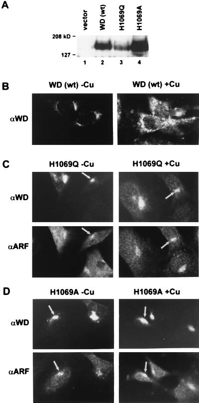
Figure 4.
(A) Immunoblot analysis of lysates from mottled fibroblast clones maintained at 28°C. Protein (125 μg) from cells stably transfected with vector alone (lane 1), wild-type Wilson cDNA (lane 2), or Wilson cDNA mutants (lanes 3 and 4) were separated by SDS/7.5% PAGE, transferred to nitrocellulose, and analyzed by chemiluminescence. (B) Immunofluorescence on wild-type Wilson clone 4 with and without copper treatment by using the Wilson protein antibody. (×600.) (C) Immunofluorescence on a Wilson H1069Q clone with and without copper treatment by using antibodies against the Wilson protein or ARF. Arrows indicate trans-Golgi network localization in corresponding cells. (×600.) (D) Immunofluorescence on a Wilson H1069A clone with and without copper treatment by using antibodies against the Wilson protein or ARF. Arrows indicate trans-Golgi network localization in corresponding cells. (×600.)

