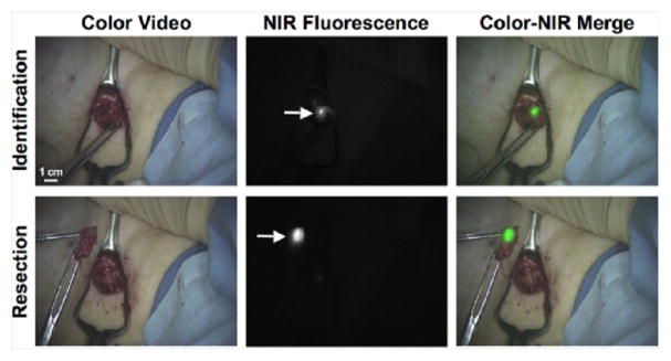
Fig. 3.
Example images from the pilot clinical trial of the FLARE™ imaging system during SLN mapping for breast cancer. The FLARE™ system collected color video and 800 nm NIR fluorescence images simultaneously. The NIR fluorescence image was pseudo-colored and merged with the color image in real-time, permitting the surgeon to resect the SLN (arrow) under fluorescence-guidance.

