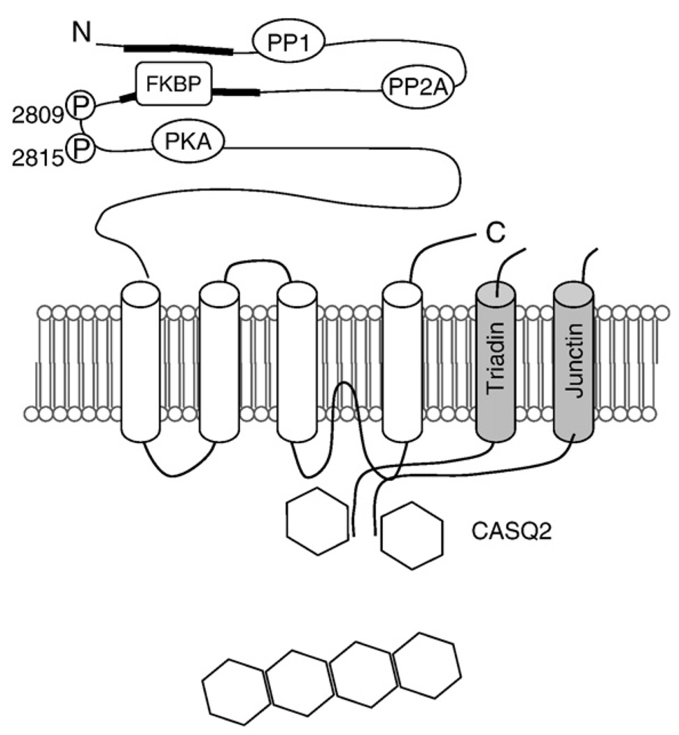
Fig. 2.
Schematic presentation of the RyR2 channel complex. The domain structure of RyR2 with sites of interaction with auxiliary proteins and phosphorylation sites. Two major divergent regions located in the cytosolic portion are indicated. Calsequestrin, junctin, and triadin, proteins interacting with RyR and between themselves and RyR2 in the SR, are shown. PP, protein phosphatase; P, phosphorylation sites; CaM, calmodulin; CaMKII, calmodulin-dependent protein kinase II. Adapted from Gyorke and Terentyev, 2008.

