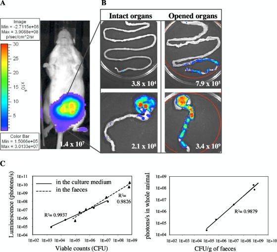
FIG. 2.
Monitoring of intestinal colonization by bioluminescence imaging in whole animals. (A) E. coli MG1655-Strr/pAL2(pVA838ΩPamiluxABCDE) (107 CFU) was inoculated intragastrically into four streptomycin-treated mice, and the bioluminescent signal was measured transcutaneously in whole animals 24 h postfeeding. The intensity of the transcutaneous photon emission is represented as a pseudocolor image. (B) The small (top) and large (bottom) intestines were dissected at 72 h, and the photon/s per organ was quantified on intact (left) or longitudinally opened (right) organs. (C) Linear correlation between the bacterial counts and the bioluminescent signal, determined by serial dilutions of bioluminescent bacteria in culture medium or in feces (left). Correlation between the transcutaneous photon emission measured in the whole animals and the number of bioluminescent bacteria in the feces during colonization (right).

