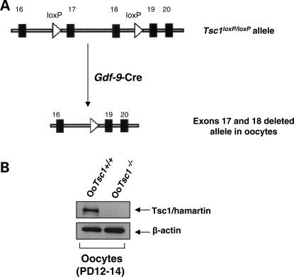
Figure 1.
Oocyte-specific deletion of Tsc1 in mice. (A) Schematic representation of deletion of Tsc1 exons 17 and 18 by Gdf-9-Cre-mediated recombination in oocytes. (B) Western blots showing the absence of Tsc1 (hamartin) protein expression in oocytes of OoTsc1−/− mice. Oocytes were isolated from ovaries of 12- to 14-day-old OoTsc1+/+ and OoTsc1−/− mice as described in Materials and Methods. For each experiment, material from 3–5 mice was used per lane. For each lane, ∼20 µg of protein was loaded. Levels of β-actin were used as internal controls. The experiments were repeated three times and representative images are shown.

