Figure 4.
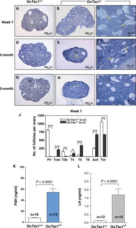
Follicle depletion and POF at young adulthood in OoTsc1−/− mice. (A–I) Morphological analysis of ovaries from OoTsc1−/− and OoTsc1+/+ littermates at 7 weeks, 2 months and 3 months of age. Ovaries from OoTsc1−/− and OoTsc1+/+ mice were embedded in paraffin and sections of 8 μm thickness were prepared and stained with hematoxylin. When compared with ovaries of OoTsc1+/+ mice (A), OoTsc1−/− ovaries still appeared larger at 7 weeks of age (B). However, many activated follicles in OoTsc1−/− ovaries had already degenerated (C, arrows). At 2 months of age, almost all follicles had degenerated in OoTsc1−/− ovaries (F, arrows) and the ovaries were smaller (E) than OoTsc1+/+ ovaries (D). By 3 months of age, healthy follicular structures had completely disappeared in OoTsc1−/− ovaries (H and I). Control OoTsc1+/+ mice had normal ovarian morphology (G). CL, corpus luteum. (J) Quantification of follicle numbers in ovaries of 7-week-old OoTsc1−/− and OoTsc1+/+ ovaries. The numbers of different types of follicles per ovary (mean ± SEM) were quantified as described in Materials and Methods. In 7-week-old OoTsc1−/− ovaries, the numbers of healthy follicles were significantly reduced compared with OoTsc1+/+ ovaries. The numbers of mice used (n) and results of statistical analyses are given. **P < 0.01, ***P < 0.001. (K and L) Levels of FSH and LH in sera of 3- to 4-month-old OoTsc1+/+ and OoTsc1−/− mice. FSH and LH were measured as described in Materials and Methods. Significantly elevated levels of FSH (K) and LH (L) in adult OoTsc1−/− mice were observed. The numbers of mice used (n) and P-values are shown in the figures.

