Figure 6.
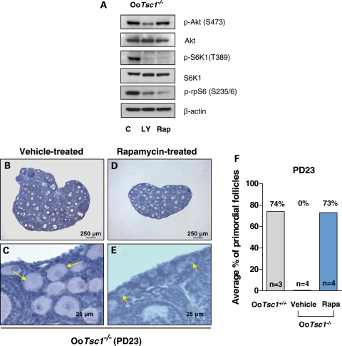
Rapamycin effectively reverses the overactivation of primordial follicles in OoTsc1−/− ovaries. (A) Activation of S6K1–rpS6 in OoTsc1−/− oocytes is dependent on PI3K and mTORC1 signaling. Oocytes were isolated from ovaries of OoTsc1−/− mice at PD12–14 as described in Materials and Methods. Treatment of oocytes with the PI3K-specific inhibitor LY294002 (LY, 50 µm) for 1 h was found to largely suppress levels of p-Akt (S473), p-S6K1 (T389) and p-rpS6 (S235/6), whereas treatment with the mTORC1-specific inhibitor rapamycin (Rap, 50 nm) for 1 h only suppressed the levels of p-S6K1 (T389) and p-rpS6 (S235/6), but not the level of p-Akt (S473) in OoTsc1−/− oocytes. This suggests that activation of S6K1–rpS6 in OoTsc1−/− oocytes is dependent on both PI3K and mTORC1 signaling. Levels of total Akt, S6K1 and β-actin were used as internal controls. (B–E) The overactivation of primordial follicles in OoTsc1−/− mice can be reversed by treatment with rapamycin. Rapamycin (5 mg/kg body weight) was injected daily into OoTsc1−/− mice from PD4 to PD22, and the ovaries were collected at PD23 for morphological analysis. Ovaries from rapamycin-treated OoTsc1−/− mice appeared smaller (D) than the ovaries from vehicle-treated OoTsc1−/− mice (B). Clusters of primordial follicles were seen in rapamycin-treated OoTsc1−/− mice at PD23 (E, arrows), whereas all primordial follicles were activated in vehicle-treated OoTsc1−/− mice at PD23 (C, arrows). (F) Proportions of primordial follicles (relative to the total number of follicles) in OoTsc1+/+, OoTsc1−/− (vehicle-treated) and OoTsc1−/− (rapamycin-treated) ovaries at PD23. The average percentages of primordial follicles are shown. The proportion of primordial follicles in rapamycin-treated OoTsc1−/− ovaries was found to be elevated to 73%, which was similar to the proportion in OoTsc1+/+ ovaries (74%). The numbers of mice used (n) are shown. Rapa, rapamycin.

