Figure 1.
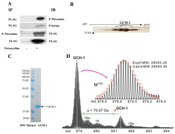
GCH-1 was phosphorylated in Flp-In 293 cells. (A) FLAG-GCH-1 cells were immunoprecipitated and immunoblotted with phospho-threonine, phospho-serine or FLAG antibodies. (B) The immunoprecipitated FLAG-GCH-1 was separated by IPG gel followed by SDS/PAGE analysis. The gel was then silver-stained and the spots were validated as GCH-1 by MS. (C) The immunoprecipitated FLAG-GCH-1 was separated by SDS/PAGE and visualized by coomassie staining. (D) High resolution ESI/FTMS analysis of GCH-1 overexpressed in HEK293 cells suggesting it is monophosphorylated. Inset, isotopically resolved GCH-1 intact protein ions of +32 charge state (M+32). Circles represent the theoretical isotopic distribution of the GCH-1 protein ions with an acetylation. pGCH-1, monophosphorylated GCH-1. +Na/+K, sodium/potassium adducts of GCH-1 protein ions. Expt’l MW, experimental most abundant molecular weight; Calc’d MW: calculated most abundant molecular weight.

