
Figure 2.
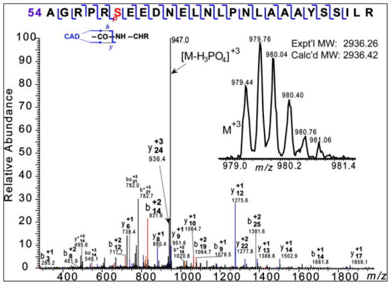
Confirmation of a phosphorylation site at S59 of FLAG-GCH-1 (corresponding to site S51 in endogenous GCH-1). CAD spectrum of an isolated +3 charge state of a tryptic peptide S[54–59]R at m/z 979 (inset), corresponding to a monophosphorylated A[54–79]R with a phosphorylated site at Ser59 (pS). Expt’l MW, experimental most abundant molecular weight; Calc’d MW: calculated most abundant molecular weight.