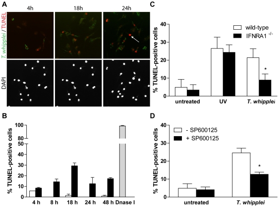
Figure 6. T. whipplei-induced apoptosis depends on type I IFN through JNK activity.
(A) BMDM were infected with T. whipplei (MOI 50∶1) for 4 h and incubated for the indicated times. Apoptotic cells were revealed by TUNEL reaction, while T. whipplei organisms were revealed using a T. whipplei specific antibody. Nuclei were stained with DAPI. (B) Apoptosis was quantified by examining 3 to 5 fields per condition (100 to 300 cells each). The percentage of TUNEL-positive cells was calculated as the ratio between TUNEL-positive and DAPI-stained nuclei ×100 (white bars: uninfected cells, black bars: T. whipplei-infected cells, gray bar: DNaseI-treated cells). (C) BMDM from wild-type and IFNAR1−/− mice were infected with T. whipplei (MOI 50∶1) for 4 h and incubated for 18 h. As a control, cells were exposed to UV. Apoptosis was determined as the percentage of TUNEL-positive cells. (*, P<0.05). (D) BMDM were treated or not with the JNK specific inhibitor SP600125 for 30 min. Cells were then infected with T. whipplei (MOI 50∶1) for 4 h and incubated for 18 h. Apoptosis was determined as the percentage of TUNEL-positive cells. (*, P<0.05).

