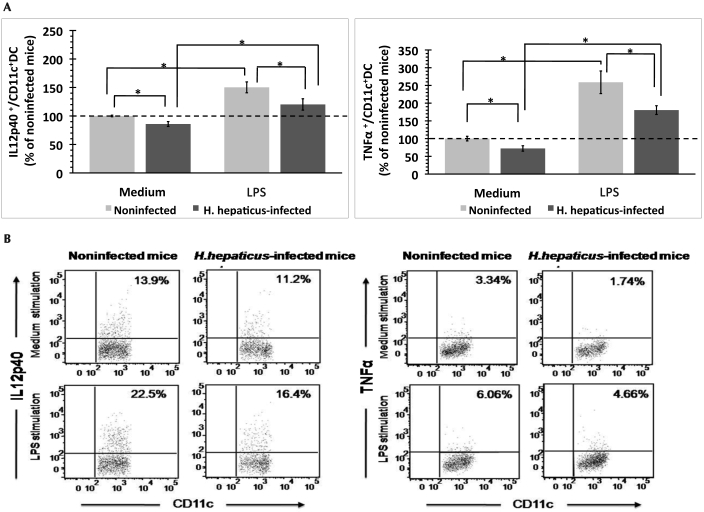
Figure 5.
Intracellular proinflammatory cytokine expression by DC in the colic LN of H. hepaticus-infected and noninfected mice after in vitro exposure to medium or lipopolysaccharide (LPS). (A) The percentage (mean ± SE) of cytokine-producing DC was normalized to the percentage of cytokine-producing cells from noninfected mice after incubation with medium within each experiment (n = 5 or 6 mice per group). Results were combined from 3 independent experiments. (B) Representative dot plots depicting the intracellular staining profile for IL12p40 and TNFα in CD11c+ DC population. *, significant difference (P < 0.05) between indicated groups.

