Figure 1.
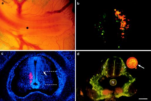
Experimental paradigm. (a) This in ovo photograph shows a bead in place on a somite 24 hours after bead placement (caudal is to the right). (b) Retrograde labeling of preganglionic neurons in T6 at E10. Preganglionic neurons that project rostrally along the sympathetic chain are labeled red, and those that project caudally are labeled green. This 30-μm section is taken from an experimental animal in which the left side is the control and the right side was treated with disulfiram. (c) Location of T6 sympathetic preganglionic neurons at E7. Sympathetic preganglionic neurons on the left side were labeled retrogradely (red) from the T6 sympathetic chain; sections were stained with Hoechst dye to label all of the nuclei. Counts of BrdU-labeled cells were made from a restricted region of the spinal cord equivalent to the area containing preganglionic neurons. The dorsoventral extent of the counted region extends from the bottom of the central canal to the point at which the ventricular neuroepithelium fans out laterally (marked by dotted lines). The mediolateral extent of the counted region extended from the edge of the ventricular epithelium to the most lateral extent of the ventricular epithelium (arrow). To determine the percentage of nuclei in this area that represent preganglionic neurons, nuclei and retrogradely labeled preganglionic neurons were counted in this restricted region in four sections such as these from five different preparations. (d) Development of the somites and migration of neural crest and formation of sympathetic ganglia were apparently normal after all treatments. Here, migrating neural crest (green fluorescence) is shown at somite 26 20 hours after treatment with a citral-containing bead (arrow). Coalescing dorsal root ganglia are marked by asterisks. n = notochord. (Calibration: a and b, 200 μm; c, 100 μm; d, 50 μm.)

