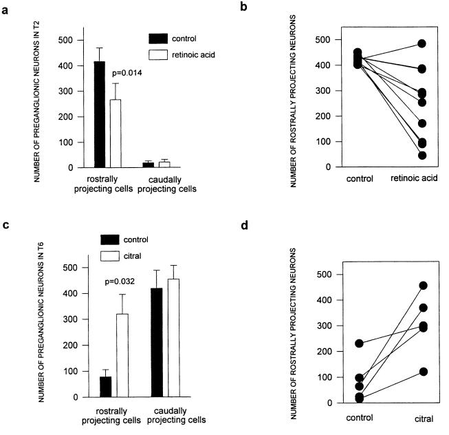
Figure 2.
Effect of altering retinoid signaling on sympathetic preganglionic projections. In contrast to Table 1, the data plotted in this figure are restricted to those cases in which the bead was placed on the penultimate somite and was recovered at T2 or T6 only. (a) Treatment of T2 somites with RA selectively decreases the number of preganglionic neurons that project rostrally. Values are mean ± SEM; significance was assessed with Wilcoxon Signed Rank test, n = 10. In 9 of 10 cases, the experimental side contained fewer rostrally projecting neurons than the control side. (b) Same data as in a, following a normalization procedure in which we normalized all data points to the average number of labeled neurons (both rostrally and caudally projecting) for the control side. The normalized data are presented in matched pairs, thus illustrating the range and magnitude of the experimental effects. (c) Treatment of T6 somites with citral selectively increases the number of preganglionic neurons that project rostrally. Data presented as in a (n = 5). In all five cases, the experimental side contained more rostrally projecting neurons than the control side. (d) Same data as in c, following the normalization procedure described in b.

