Fig. 2.
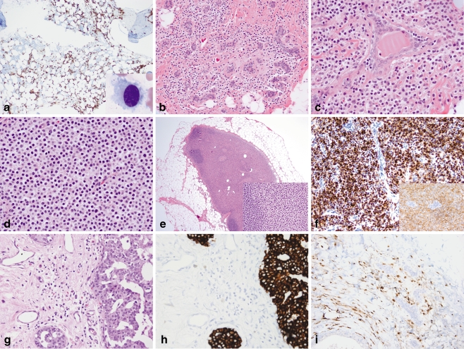
Hairy cell leukemia involving the bone marrow, breast, and axillary sentinel lymph node. a A posterior iliac crest bone marrow aspirate smear contains medium-sized lymphoid cells with oval nuclei, dispersed chromatin, and abundant pale cytoplasm with circumferential cytoplasmic projections (inset, Wright-Giemsa, ×1,000). CD20 stain of the core biopsy highlights the interstitial pattern of marrow involvement characteristic of hairy cell leukemia (×100). b–d Left breast lumpectomy containing prominent monomorphic infiltrates of small discohesive cells with abundant pale cytoplasm and round to oval nuclei present singly and in linear strands surrounding benign lobules (b; H&E, ×200), as larger clusters and aggregates surrounded by thin bands of fibrous stroma (c; H&E, ×400), as well as in solid sheets (d; H&E, ×400). DCIS was present elsewhere (not shown). e The sentinel lymph node shows massive replacement of the paracortex and hilum by cells with abundant pale eosinophilic cytoplasm and oval to spindled nuclei, morphologically similar to those in the breast (inset, H&E, ×400); residual primary follicles are present at the periphery of the node (H&E, ×20). f Immunohistochemistry of the breast specimen reveals infiltrating cells to be CD20-positive (inset) and strongly DBA.44-positive (×400). g Subsequent mastectomy demonstrates an infiltrate of monomorphic cells present singly and in loose linear arrays adjacent to residual cribriform DCIS (H&E, ×400). h–i A cytokeratin cocktail immunostain (h) of the mastectomy specimen highlights DCIS, but not the infiltrating cells, which are positive for CD20 (i; ×400)

